Eman Abu-Gharbieh1,2 ![]() ,
Naglaa G Ahmed3,4
,
Naglaa G Ahmed3,4
For correspondence:- Eman Abu-Gharbieh Email: eabugharbieh@sharjah.ac.ae Tel:+97165057289
Received: 24 May 2016 Accepted: 15 September 2016 Published: 31 October 2016
Citation: Abu-Gharbieh E, Ahmed NG. Bioactive content, hepatoprotective and antioxidant activities of whole plant extract of Micromeria fruticosa (L) Druce ssp Serpyllifolia F Lamiaceae against Carbon tetrachloride-induced hepatotoxicity in mice. Trop J Pharm Res 2016; 15(10):2099-2106 doi: 10.4314/tjpr.v15i10.7
© 2016 The authors.
This is an Open Access article that uses a funding model which does not charge readers or their institutions for access and distributed under the terms of the Creative Commons Attribution License (http://creativecommons.org/licenses/by/4.0) and the Budapest Open Access Initiative (http://www.budapestopenaccessinitiative.org/read), which permit unrestricted use, distribution, and reproduction in any medium, provided the original work is properly credited..
Purpose: To investigate the antioxidant and hepatoprotective activities of Micromeria fruticosa Druce (L.) Druce ssp Serpyllifolia F. Lamiaceae (MF) extract and to correlate its phenolic composition of the biological activities.
Methods: Reversed-phase high-performance liquid chromatography (RP-HPLC) was employed for the identification and quantification of phenolics. 2,2-Diphenyl-1-picrylhydrazyl (DPPH) radical scavenging potential of the four extracts, namely, ethanol, methanol, acetone, and ethyl acetate, were assessed. The hepatoprotective and antioxidant activities were evaluated against carbon tetrachloride (CCl4)-induced hepatotoxicity in mice. Antioxidant status in the liver was assessed by determining the activities of some antioxidative enzymes, namely, superoxide dismutase (SOD), catalase (CAT) and glutathione peroxidase (GSH-Px), and the levels of thiobarbutaric acid reactive substances (TBARS).
Results: RP-HPLC analysis revealed high contents of quercitrin, rosmarinic and ferulic acid. The four extracts were potent DPPH free radical scavengers. Administration of the ethanol extract to the animals twice daily for 14 days did not show any evidence of hepatotoxicity. CCl4 caused a marked increase in TBARS and significant decrease in CAT, GSH-Px and SOD levels, but this was reversed by the ethanol extract.
Conclusion: The ethanol extract of Micromeria fruticosa (L) may have a palliative effect in liver injuries and this is probably due to the antioxidant properties of the plant’s polyphenolic content.
Introduction
Phenolics are plant metabolites with well-known protective action against various health diseases [1]. For instance, they possess numerous biological activities e.g. anti-inflammatory [2], anti-diabetic [3], antioxidant, cytotoxic and antitumor [4,5].
Micromeria fruticosa Druce (Wildflowers, White Micromeria) is used widely in many Mediterranean countries as herbal infusion for various inflammatory conditions and in wound healing [6]. It is a member of genus Micromeria and is known as zutalevana and ashab a-shai in Hebrew and Arabic respectively. The essential oil of Micromeria fruticosa (MF) is comprised largely of monoterpenes (+)-pulegone and isomenthol [7;8]. The aqueous extract of the plant is reported to possess anti-inflammatory [6;7], analgesic [8] and gastroprotective activities [6]. Also, the aqueous extract and the volatile oil exhibited marked antitumor activities against human colon tumor cells and mammary carcinoma F7 [8]. Toxicity study of its aqueous extract was reported previously on mice and it was safe up to 5 g/kg [8].
The current study aimed to evaluate the possible hepatoprotective and antioxidant activities of the ethanol extract of Micromeria fruticosa, and to determine if there is a correlation between its phenolic composition and biological activities.
Methods
Chemicals and drugs
Chloroform, ethanol, methanol, acetone, and ethyl acetate were purchased from Fisher Scientific, USA. Carbon tetrachloride (CCl4), 2,2-diphenyl-1-picrylhydrazyl (DPPH), sodium carboxymethylcellulose (CMC), biochemical kits for determination of glutathione peroxidase (GPx), superoxide dismutase (SOD), catalase (CAT) and thiobarbituric acid reactive substances (TBARS) were purchased from Sigma Chemical Co. (St. Louis, MO, USA). Folin-Ciocalteu reagent was obtained from Merck (Darmstadt, Germany). All other chemicals were of analytical grade.
Plant material
The whole plant of Micromeria fruticosa was collected in July 2012 from Nablus, Palestine. In August 2012, the sample was identified and authenticated by Dr. Hassnaa Ahmed Hosny, Professor of Plant Taxonomy, Department of Botany, Faculty of Science, Cairo University, Egypt. Voucher specimen was kept at the Herbarium of Dubai Pharmacy College, Dubai, UAE. Sample, air-dried in shade, powdered and preserved for the current study.
Experimental animals
Thirty healthy male albino mice (30 - 35 g) were used. The animals were kept under the same standard conditions (temperature 22.0 ± 2.0 ºC, relative humidity 50 - 60 %, with 12 h day/night light cycle), fed with well-balanced normal diet purchased from LabDiet® and water supplied ad libitum. All mice were acclimated for one week before commencing the experiments. All the animals were treated according to the Guide for the Care and Use of Laboratory Animals published by the US National Institutes of Health [9]. The experimental procedures were approved by the Research Ethical Committee of the Dubai Pharmacy College, Dubai, United Arab Emirates (ref no.14-11-2012)
Plant extract preparation
The air-dried powdered plant material (1 kg) was exhaustively extracted twice by cold maceration in five liters of 70 % ethanol. The solvent was evaporated under reduced pressure at 50 oC to yield 200 g dry residue. The dried extract was kept in a refrigerator and used for biological evaluation study.
Assay of phenolic content
Solvents of different polarities, namely 70 % ethanol, methanol, acetone and ethyl acetate, were individually used for extraction of the air-dried powdered plant material (100 g each) through cold maceration. The solvents were separately evaporated under reduced pressure at 50 oC. Total phenolic and flavonoid contents were colorimetrically estimated by spectrophotometer (UV-1700 Pharma Spec, Shimadzu, Japan). The experiments were carried out in triplicate.
Total phenolic content was assessed by Folin-Ciocalteu method described by Oktay et al [11]. Results were expressed in mg/g gallic acid equivalent, calculated on dry weight of plant material; serial dilutions of gallic acid (10, 20, 30, 40 and 50 𝜇g/mL) were used for establishment of the calibration curve.
Aliquot (1 mL each) of test samples and standard was, separately, added to 9 mL of water in a volumetric flask followed by 1 mL of Folin-Ciocalteu reagent. Furthermore, the reaction mixture was vortexed and left to stand for 5 min. Thereafter, 10 mL of 7 % sodium carbonate was added and the mixture was incubated for 90 min, at room temperature. Finally, the absorbance was measured at 750 nm against the reagent blank.
Total flavonoid content of the extract was measured spectrophotometrically by the aluminum chloride method, using quercetin as standard [12]. Plant extracts (0.1 mL each) were mixed individually with 0.3 mL distilled water and 0.03 mL of 5 % sodium nitrite. The reaction mixture was left for 5 min, at 25 ∘C. Aluminum chloride solution (0.03 mL, 10 %) was added, and the mixture left for another 5 min. Later, NaOH solution (0.2 mL, 1 mM) was added to the mixture, and diluted with distilled water up to 1 mL. Absorbance was measured at 510 nm.
RP-HPLC analysis of phenolics
The phenolic composition of the methanol extract of MF was investigated in aliquots (1 g,) with RP-HPLC on a Hewlett Packard HPLC System (HP 1050 HPLCDADw/Data System). Analyses were carried out at operating conditions suitable for detection of both phenolic acids and flavonoids [13;14]. For determination of phenolic acids, the apparatus was equipped with an Alltima C18 column (particle size 5 mm, 150 × 4.6 mm) and Alltima C18 guard column (5 mm) (Alltech, USA), the UV detector being set at 280 nm. In addition, the separation of flavonoids was carried out on a Hypersil-ODS C18 column (particle size 5 𝜇m, 4.6 × 250 mm) and the UV detector was set at 330 nm. All analyses were performed at 35 oC; gradient elution was employed using acetonitrile-acetic acid mixtures as mobile phase, at a flow rate of 1 mL/min, and the injection volume was 10 𝜇L for both standard and test samples. Authentic reference samples were prepared by diluting stock solutions with methanol to prepare a final concentration of 50 𝜇g/mL. Identification of individual components was performed by comparing their retention times with those of the available standards similarly analyzed. Quantification was based on peak area computation using the external standard method. All analyses were carried out in triplicate and the results were recorded as mean.
2, 2-Diphenyl-1-picrylhydrazyl (DPPH) radical scavenging assay
Different solvents were used for the extraction of MF namely, 70 % ethanol, methanol, acetone, and ethyl acetate. All the extracts were tested in vitro for their abilities to scavenge the free radicals by using the stable DPPH radical. The assay was performed in a 96-well microtiter plate using a previously described method [15].
A percentage inhibition (%) of the DPPH radical by the samples was calculated using Eq 1.
Inhibition (%) = {A0 - (A1- A2)/A0} x 100 …….. (1)
where A0 is the absorbance of the control, A1 is the absorbance in the presence of the sample and A2 is the absorbance of the sample under identical conditions as A1 with ethanol instead of DPPH solution. Ascorbic acid (AA) was used as a reference compound. IC50 values were calculated. Samples were analyzed in triplicate.
Evaluation of hepatoprotective effect
The rats were randomly assigned to five groups of 6 animals each. The residue of ethanolic extract was suspended in 1 % CMC.
Group I: served as normal control and received the vehicle orally (1 % CMC) during the experiment period. Group II: served as a hepatotoxicity control group, and animals received 1 % CMC solution orally for 14 days. Three groups were devoted for MF extract treatment; group III: received the plant extract orally twice daily at a dose of 200 mg/kg for 14 days to assess the possible hepatotoxicity that may arise from by the plant itself. Groups IV and V were used to evaluate the possible hepatoprotective activity of the plant extract. Thus, animals were pre-treated with the extract for 14 days in two different doses (100 and 200 mg/kg) respectively. After a 14-day treatment, hepatic injury was induced by intraperitoneal injection of 1.0 ml/kg of CCl4 in groups II, IV and V and the mice were sacrificed six hours after the last treatment by ether anesthesia.
Liver enzyme assessment
Blood samples were collected from the animals’ hearts individually with the use of 3 ml sterile syringe and blood was transferred immediately into non-heparinized tubes and used later for the analyses of liver enzymes: alanine aminotransferase (ALT), aspartate aminotransferase (AST) and alkaline phosphatase (ALP).
Assay of antioxidant enzymes in liver homogenate
Liver samples were removed surgically from the mice immediately and kept in -80 οC in So-Low Ultra Low Upright Freezer for further antioxidant enzyme assay. The activity of catalase, superoxide dismutase, glutathione peroxidase and TBARS was assessed as per the method described by Abu-Gharbieh et al [16].
Histopathological examination
Samples from the liver were suspended in 10 % formaldehyde for further histological assessment. Liver tissue was processed and embedded in paraffin wax. Sections of 5 μm in thickness were cut and stained with hematoxylin and eosin (H & E) stain and assessed under a light microscope (CH20-Olympus®).
Statistical analysis
The data are reported as mean ± standard deviation (SD, n = 3). The data were analyzed using one-way analysis of variance (ANOVA), followed by Dunnett’s multiple comparison test (DMCT). P < 0.05 was considered statistically significant.
Results
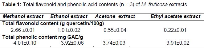
Influence of extracting solvent on total phenolic and flavonoid contents
All the tested extracts were found to be rich in both phenolic acids as well as flavonoids compounds as shown in . The highest amount of flavonoids was detected in the methanolic extract (2.66 %) followed by ethanolic extract (1.01 %). Ethyl acetate and acetone extracts showed flavonoid content of 0.55 and 0.22 % respectively. On the other hand, all the tested extracts showed approximately similar content of phenolic acids calculated as mg/g gallic acid.
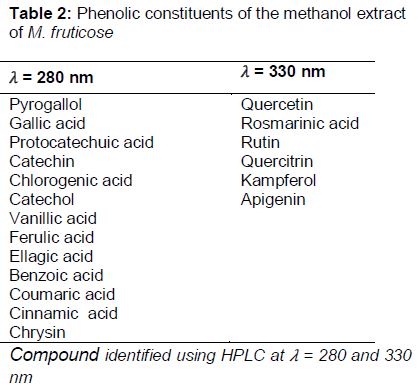
Phenolic content
A total of 13 components (corresponding to 22.83 % of the total composition) were identified at 280 nm as shown in . Among them, eight phenolic acids were identified (13.69 %) with prevalence of ferulic acid (4.30 %). Moreover, two flavonoids, catechin and chrysin, and a diphenol compound, namely catechol were detected. A non-phenolic compound, benzoic acid, was present in a high concentration (2.08 %). On the other hand, by setting the detector at 𝜆 = 330 nm, six compounds were identified (). Among them, five were flavonoids with major quercitrin (4.72 %). Moreover, rosmarinic acid (phenolic acid) was detected in a high amount (4.23 %).
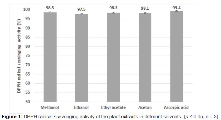
DPPH free radical scavenging activity
The four MF extracts were found to be potent DPPH radical scavengers comparable to the ascorbic acid as shown in . No statistical differences were found between the tested extract groups and the reference (p < 0.05).
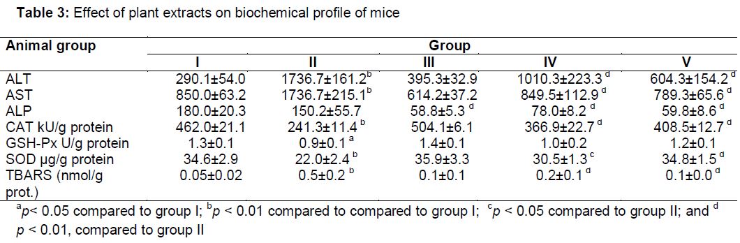
Serum markers of liver damage
Injection of carbon tetrachloride induced a significant increase of ALT and AST by six and two folds respectively compared to control group (p < 0.01) , while ALP was not significantly affected as shown in .
Administration of MF extract twice daily over two weeks did not show any evidence of hepatotoxicity, as serum ALT and AST levels did not significantly change (). A significant reduction in ALP level was noticed.
Injecting carbon tetrachloride to the animals pretreated with the plant extract at two different doses (100 and 200 mg/kg) resulted in elevation of both ALT and AST levels in serum, this elevation was significantly lower than that caused by CCl4 alone. Noticeable effect occurred in a dose dependent manner since the p-values were less than 0.05 and 0.01 for the 100 and 200 mg/kg respectively compared to group II. Furthermore, ALP level was reduced significantly in MF treated mice.
Histopathological findings
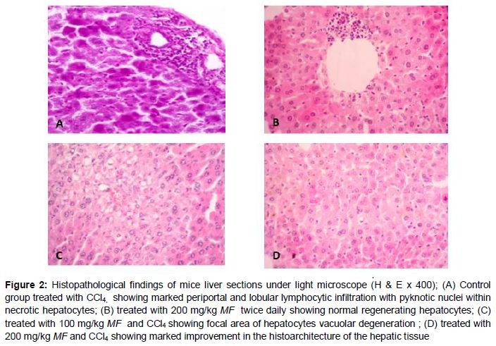
Histopathological analysis revealed a dense periportal and lobular lymphocytic infiltrate with diffused pyknotic nuclei within necrotic hepatocytes in periportal areas in the liver tissue obtained from the control group treated with CCl4 alone (A). On the other hand, gross hepatocyte renewal and regeneration were observed after the administrating the MF extract twice daily for two consecutive weeks as shown in Figures 2B. Furthermore, pretreating the animals with the plant extract at two different doses improved the histoarchitecture of the hepatic tissue in a dose dependent manner as shown in Figures 2C and D.
Effect of MF on antioxidant enzymes and TBARS contents
Administration of CCl4 markedly depleted the antioxidant enzymes (CAT, GSH-Px and SOD) in the mice livers (). Nevertheless, CCl4 increased significantly (p < 0.01) the hepatic lipid peroxidation that is expressed by high TBARS content.
Giving the plant extract twice daily for two weeks showed slight enhancement of the antioxidant enzymes and mild reduction in the TBARS content, but they were all statistically insignificant as shown in .
Pretreating the mice with the plant extract at different doses reversed significantly the reduction in catalase and superoxide dismutase levels caused by CCl4. On the other hand, the highest dose increased the glutathione peroxidase level but this increment was statistically insignificant. Moreover, MF treatment, at both doses, was effective in inhibiting the hepatic lipid peroxidation, represented by the TBARS content.
Discussion
Liver is the main organ involved in generation of reactive oxygen species (ROS) induced by various drugs and chemicals [19]. It is well-known that hepatotoxicity involves induction of oxidative stress due to excessive formation of ROS which causes extensive lipid peroxidation of the hepatocyte cellular membranes [20,21].
Various medicinal plants with long and well-established traditional uses have been recommended to treat several hepatic disorders [22]. Yet, clinical application of those plants needs extensive in vivo pharmacological and toxicological characterization, in addition to extensive clinical studies to explore their usefulness in such diseases.
ALT is found predominately in the liver, with lesser quantities found in the kidneys, heart, and skeletal muscle. As a result, the ALT is a more specific indicator of liver inflammation than AST, as AST may also be elevated in diseases affecting other organs, such as the heart or muscles. Elevation of the alkaline phosphatase level is usually associated with cholestatic liver diseases [23].
Micromeria fruticosa is used widely in many Mediterranean countries for its traditionally known health benefits. It is used commonly to heal wounds and in various inflammatory conditions [6]. In literatures, the aqueous extract of Micromeria fruticosa is reported to show many pharmacological actions such as anti-inflammatory [6;7], analgesic [8] and gastroprotective activities [6].
Although the plant extract was not previously investigated for its bioactive components, its oil was analysed and it was found to contain high amount of oxygenated compounds [8]. The present study investigated the bioactive components of the plant extract, especially phenolics. Phenolics are considered the medicinal treasure for any plant, since they possess various biological activities such as anti-inflammatory, antidiabetic, anticancer and antioxidant [2-5].
Quantitative determination of total phenolics revealed that all extracts were rich in phenolic acids and flavonoids, but the highest amount of flavonoids was present in the methanol extract followed by ethanol extract.
DPPH assay was applied as in vitro approach to assess the free radical-scavenging activity and to screen for the possible antioxidant activity of the different plant extracts. The assay showed that all plant extracts had strong DPPH radical scavenging activity comparable to those of ascorbic acid regardless the type of the extraction solvent. However, for safety, availability and economic concerns, the ethanol extract was selected for the biological study.
Dose selection for the subsequent biological study for the plant extract was done based on its LD50 value (less than 1/10 of LD50). Moreover, similar doses of Micromeria fruticosa were tested previously for its anti-inflammatory, gastropro-tective [6] and analgesic [8] activities.
Administration of the plant extract for two weeks enhanced hepatic regeneration and maintained normal liver function. Furthermore, possibility of cholestasis was ruled out since ALP level remained low [24].
The hepatoprotective effect of MF extract had been evaluated on carbon tetrachloride induced hepatotoxicity model in mice. The data showed that injecting the animals with CCl4 resulted in severe liver damage evidenced by sharp increase in hepatic lipid peroxidation and both ALT and AST. Moreover, administration of CCl4 to the animals caused extensive hepatic oxidative stress that is characterized by significant decrease in CAT, GSH-Px and SOD enzymes levels and increase of TBARS content in liver tissue. Furthermore, histopathological assay revealed severe structural loss of hepatic tissue distinctive by dense periportal and lobular lymphocytic infiltrate with diffused pyknotic nuclei within necrotic hepatocytes.
Pre-treating the animals with MF extract for two weeks at two different doses (100 and 200 mg/kg) reduced the intensity of liver damage induced by carbon tetrachloride. ALT and AST levels were also elevated following CCl4 administration. This elevation was significantly less than that caused by CCl4 alone. ALP serum concentration continued to be low in all plant treated groups, that role out cholestasis. Furthermore, the extract reduced significantly the oxidative stress induced by CCl4 and it enhanced the hepatic antioxidant defense system that is represented by CAT, GSH-PX and SOD enzymes and was highly efficient in reducing TBARS content in liver tissue in a dose related manner.
These findings were supported by the histological results since it showed that all harmful effects of carbon tetrachloride on liver tissues were attenuated and tissue architecture was maintained by pretreating the animals with the plant extract in a dose-dependent manner. HPLC analysis revealed that rutin and quercetrin were the main flavonoidal glycosidal compounds present in MF extract. Those flavonoids which contain sugar moiety are most likely hydrolyzed to their aglycones after their oral administration by bacterial intestinal enzymes to quercitin [25] and then absorbed into the intestinal cells by passive mechanisms.
Quercitin has been extensively documented in literature to process high antioxidant activity. Due to this metabolic process of the glycosides, quercitin content available in the intestine appears to be much higher than that present in HPLC data. Accordingly, the hepatoprotective effect of the plant extract could be related to the higher content of different groups of phenolic compounds including flavonoids and phenolic acids that have been earlier reported to exhibit strong antioxidant and hepatoprotective effects.
Conclusion
Micromeria fruticosa extract effectively protects against the hepatotoxicity induced by CCl4 through scavenging of free radicals in mice as well as by boosting the antioxidant capacity of the liver, probably by the bioactive antioxidant principles present in the extract. Therefore, the plant is a potential safe source of protective agents against liver damage.
Declarations
Acknowledgement
References
Archives


News Updates